Beam size estimates made using beam-beam deflections
are used for optimization of the Stanford Linear Collider (SLC)
electron-positron beam sizes. Beam size and intensity goals for
1996 were 2.1 x 0.6 µm (x,y) at 4.0x10 10 particles
per pulse. Conventional profile monitors, such as scanning wires,
fail at charge densities well below this. Since the beam-beam
deflection does not provide single beam information, another method
is needed for Interaction Region (IP) beam size optimization.
The laser based profile monitor uses a finely focused 349 nm.
wavelength , frequency-tripled YLF laser pulse that traverses
the particle beam path about 29 cm away from the e+/e- IP. Compton
scattered photons and energy degraded e+/e- are detected as the
beam is steered across the laser pulse. The laser pulse has a
transverse size, ( s0,
), of 380 nm and a Rayleigh range of about 5 µm. This is
adequate for present or planned SLC beams. Design and results
are presented.
The Stanford Linear Collider (SLC) is the first of a new generation of colliding beam machines that rely on micron sized beams colliding at a relatively low repetition rate []. The ultimate performance of the collider is limited by the control of emittance dilution effects that increase the transverse beam size (sx,y) at the interaction point (IP), where the two beams meet each other. A useful estimate of sx,y is obtained from the deflection seen when sweeping one beam across the other []. The two principle drawbacks of the deflection technique are its sensitivity to shifts in beam centroid and a lack of indication of which beam is changing.
The latter has the most significant impact on emittance dilution and optics related optimization. A single beam (e+ or e-) diagnostic that can operate over the full range of SLC beam intensities from 0.3 x 10¹⁰ to 4 x 10¹⁰ particles per bunch is needed. Unfortunately, wire scanners cannot be used for beam sizes smaller than 1.4 µm or for intensities greater than 0.6 x 10¹⁰. A wire scanner equipped with 4 µm diameter carbon wire is used to measure sx,y for beams safely away from these thresholds. If either threshold is exceeded, the energy deposited in the wire from a single pulse severs it. The wire scanner is installed deep inside the solenoidal SLC Large Detector (SLD) system and is virtually inaccessible, so routine replacement of the carbon wires is not practical.
The SLC IP laser based beam profile monitor will be used to measure sx,y of individual beams inside the SLD over the full range of operating intensities with about 10% accuracy. Some key features of the device are similar to the laser based beam size monitor developed for the FFTB at SLAC [][]. A finely focused laser pulse is brought into a 90_ crossing angle collision with the electron and positron beam and the Compton scattered photons and degraded beam particles are detected. As the e+/e- beams are steered across the laser pulse on a succession of pulses, the amplitude of the scattered radiation is recorded and used to estimate the beam sizes, in a manner similar to that used with SLC wire scanners [].
Future linear colliders (NLC) will employ beams of
higher charge density than those of SLC []. In most designs, conventional
wire scanner limits will be exceeded for all damped beam regions.
Sets of laser based monitors will serve as emittance monitors.
Lessons learned from the operation of the device described in
this paper will be useful for the development of NLC beam profile
monitors.
We considered three basic 'optical scattering structures', 1) a diffraction limited, finely focused waist (TEM'00' mode), 2) an interference fringe pattern similar to that used at FFTB and 3) the finely focused waist of a first order transverse mode laser beam (TEM'01' mode).
The minimum transverse size, s0,
of a diffraction limited laser beam is []:
![]() .
.
where l
is the wavelength of the light, f is the focal length of
the lens and s
is the gaussian beam sigma as defined by the photon density. The
effective length of the focused section is twice the Rayleigh
range (zR).
![]() where
where ![]() .
.
The incoming beam size, sin, and f combine to give an 'f#', with the aperture roughly ±3 sin giving s0 ~ 1/2 f# l . For typical SLC parameters, with the required beam stay clear distance of 25mm and an f# of 2, l must be shorter than 500 nm in order to have s0 < sy and zR ~ 5µm. This is the option selected.
The second 'optical scattering structure', the interference fringe pattern, is useful for much smaller beam sizes than those expected for SLC and, unless the pattern pitch is controllable, can be used to measure only a small range of beam sizes. Scanning with this system involves measuring the modulation depth of the scattered radiation as the beam is moved across the pattern. We could not find room in the confines of SLD for fringe pitch control.
The third option mentioned above, the TEM '01' mode laser beam, with a field null at the center of the spot, may prove useful. It is easy to implement since the mode would be generated at the laser and the modest increase in sin is accommodated in the transport and IP optics. A double lobed result is generated from a '01' mode scan and the spacing between the lobes can be used a laser beam diagnostic.
As the particle beam is swept over the laser beam
with a varying impact parameter y0, the number
of scattered photons, Ng(y0),
is given by:
![]()
where P is the power of the laser beam intercepted by the particle beam (sz=750µm), n is the frequency of the laser light, Ne is the number of electrons in the beam, sc is the Compton scattering cross section and ss is the overlap size (ss2 =sy2 + s02 ). For peak laser power of 10MW and with sy = 1µm and Ne =1010, Ng is ~5000. A correction, nominally about 12%, is required since the particle beam has an aspect ratio (sy/sx) ~ 5 and the laser spot does not have a ellipsoidal cross section.
The energy distribution of the scattered photons
and degraded beam particles is relatively flat and, for l=350
nm (3rd harmonic YLF), has a peak gamma radiation energy of 29GeV
for SLC. Detectors for monitoring the gamma rays and the degraded
e+/e- particles are located along the beamline that extends to
the beam dump area [],[]. Backgrounds in the degraded particle
detectors are typically about 50 particles per beam crossing.
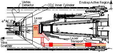
Optics design goals were to achieve the minimum s0 with the highest transmitted power and the lowest possible aberrations. The most important mechanical constraints were the minimum beam transport diameter of 25mm, the available beamline length of 52mm and mass and density restrictions. Figure 1 shows an elevation view of the IP layout. Within the cone subtended by active SLD segments, the mass of the optics and related supports must cause minimal scattering of the decay particles the SLD is intending to analyze.
The IP f2 optics (figure 2) are catadioptric, with minimal geometric aberrations, in which a diverging meniscus lens is coupled with a spherical reflector []. Two such systems are required, for measuring both sx and sy. For laser pulse lengths longer than about 150ps, the particle beam will also scatter some of the incoming photons before they reflect from the spherical mirror.
The incoming laser beam has sin of about 2.5mm. As sin is increased, diffraction scattering from the edges of the input optic produces non-gaussian tails effectively increasing s0. For small sin, the effective f# of the IP is increased, resulting in a larger s0. At the end of the transport, in the IP 'optics bench' (fig. 3), a compact switch system is used to select which of the two possible paths through the IP the laser will follow. A brewster polarizer is used in conjunction with a linear polarizer at the laser to do the selection. A compact construction was required in order to minimize the mass obscuring the SLD end cap detector segments and in order to fit around SLD internal masking and supports.
An estimate of the deviation from diffraction limit
caused by surface figure distortions yielded a per element mirror
and lens surface figure tolerance of l/40.
Since we require reliable operation for several years, about 100 million pulses, and since much of the system is sealed and inaccessible, considerable design effort was concentrated on damage prevention. In order to prevent inadvertent high power density related damage, no lenses or other focusing elements are present in the transport line that carries the light from the laser room to the IP.
The most threatening source of damage is from chemical contaminants. Studies of damage in sealed laser systems (typically infrared lasers) have shown that organic chemical contaminants are a leading cause[]. Trace contaminants, such as silicone sealers, have been pinpointed as root causes. There is little information available concerning the long term operation of UV lasers where it is possible that the sensitivity to trace organic chemicals will be greater. For this reason, our design transport s is as large as possible and all IP, IP bench and transport assembly took place in class 100 clean rooms. Non-ultra high vacuum (UHV) volumes are purged with an Ar/O2 (90/10) mixture as suggested in the report noted above.
Bulk multi-photon damage is another serious concern
for long term operation of the UV system with several transmissive
optics. Darkening in fused silica has been seen following long
term exposure to high fluence, short wavelength light []. We interpolated
between results at 248nm and at 550nm and set our tolerance a
factor 5 away from the threshold where these effects are first
seen.
Three subsystems, the IP and its mirror bench, the 17m transport line and the laser itself comprise the profile monitor. The laser is housed in a external clean room. The transport line passes through the beamline radiation shielding and then along the superconducting quadrupole triplet before entering the SLD vertex area.
The beam enters the IP, (figure 2), from the right
or from the bottom, for y or x scans respectively,
and passes through the UHV fused silica window that separates
the gas filled optics bench from the beamline vacuum. A seal design
was developed that allowed the window surfaces to be ground to
l/40 surface
quality after attaching the weld ring. The window is coated with
a graded index 'Solgel' anti-reflective [] coating. Conventional
dielectric layer coatings were rejected since their application
would be made after the attachment of the weld ring.
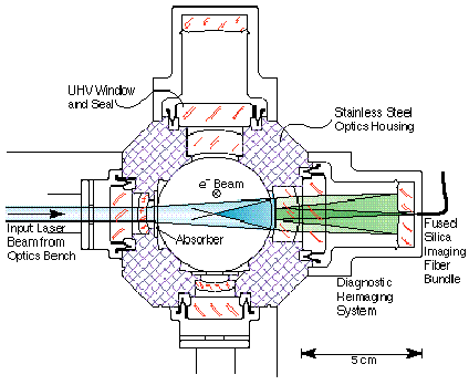
After passing through the focal point the spent laser light is absorbed using glass absorbers located both inside and outside the vacuum system. This prevents possible reflection from metal surfaces that might make secondary ghost images or sputter nearby metal surfaces and thereby cause damage to optical components. The spherical reflector coating allows 1% of the incident light to pass through. Its rear surface, together with a second spherical reflector outside of the vacuum chamber, generate an image of the IP spot that is used as a diagnostic.
The IP optics bench directs the light from the transport to the IP using one controllable and 4 fixed mirrors. It terminates the 1 inch diameter laser transport line. The transport line is evacuated for 90% of its length (10-6 Torr) and slightly pressurized, along with the optics bench, for the remaining 10%. This section is flexible, to be compliant in seismic events, and removable, to allow servicing SLD luminosity components.
The IP housing has a required vacuum performance
of 5 nano-Torr. The catadioptric optics are most sensitive to
lens and mirror centering and coplanarity errors which lead to
machining tolerances of 5µm.
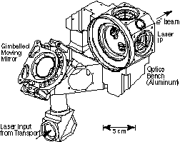
A gimbaled moving mirror mount was developed for
the transport line that has angular stability of 30µrad and
compensated bellows vacuum forces. Fine alignment adjustments
are made remotely using compact piezo-electric motors [].
Steering and alignment diagnostics are provided by 4 miniature CCD profile monitors located behind transport line and IP bench mirrors. They view the laser light directly through the partial transmission of the mirror. A frame grabber based video analysis system is used to monitor the position and size of the spot at each of the monitors.
At the monitor beyond the IP, where reliability and camera radiation damage are concerns, the 50µm spot generated by the re-imaging optics is transmitted by a 10,000 fiber fused silica bundle [] from the high radiation area to a monitor table. The coherent fiber bundle fluoresces and shifts the UV into visible. It has a diameter of 0.5mm and a single fiber diameter of 10µm. The IP assembly also has two fast diodes and a beam pickup electrode that are used for finding the relative timing of the laser and particle beams.
The transport line is protected from stray laser
light using a CW He-Cad 350 nm
l alignment laser that is mode matched to
the high power laser on the laser table. The 1mW He-Cad laser
is ideal for this purpose since it's wavelength is very close
to the tripled YLF wavelength. If the signal from the CW laser
at the monitor beyond the IP is lost, the high power laser shutter
will be closed in order to protect the transport from mis-steered
high power laser pulses.
The laser system consists of a mode-locked 119MHz, 150ps pulse length, YLF oscillator seed laser and a YLF regenerative amplifier laser. Both lasers are lamp - pumped. The oscillator is locked to the 119MHz accelerator clock frequency with a acousto-optic amplitude modulator operating at the first sub-harmonic. A single 1nJ pulse from the oscillator is switched into the amplifier using two Pockels cells.
The amplifier has a gain of 106 in 20
passes and produces 10mJ pulses of 1047nm l
light at a repetition rate of 40Hz. After tripling, we observe
2mJ at 349nm .l
with the same pulse length. We chose YLF as the lasing material
because of its reduced thermal lensing compared to YAG. After
frequency tripling, a pinhole spatial filter is used to improve
the beam shape. A small fraction of the light is diverted into
a phase space monitoring diagnostic, consisting of a transport
of roughly equal length to the main transport and a camera used
for measuring beam size, before the entrance to the transport
line. The laser beam quality, characterized by M2,
the deviation from diffraction limit, is measured to be M2=1.1.
The number of detected gamma rays and degraded e+/e- is reduced about a factor of 20 from Ng due to the finite acceptance of the detectors and transmission losses in the laser transport and IP system. Thus the expected signal at optimum overlap was about 800 degraded particles. The most difficult aspect of commissioning the monitor was establishing collisions for the first time. Laser timing and e+/e- beam x and y position must be adjusted in order to establish collisions. Since the e+/e- bunch length is short ( sz~ 2mm), the overlap is dominated by the laser pulse length. The initial timing setup done with the pickup and fast diode was accurate to about 0.5ns. The search strategy we adopted was to scan perpendicular to the laser beam direction (up and down in fig. 2) with an un-focused e- beam (one with the waist at the e+/- IP, 29cm away). A signal of a few counts over background was visible for ±1mm along the laser path. Once collisions were first established, they could easily be re-established using the timing signals and the beam position monitors that surround the IP.
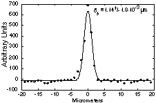
Scanning is done using controls similar to those used for the beam-beam deflection. With this system, the 120Hz beam is steered across the laser spot on a succession of pulses, with detector data recorded on each pulse. A typical background-subtracted scan is shown in figure 4. A scan like this one takes about 30 seconds to complete.
An accurate calibration procedure is required in
order to get an estimate sx,y
from the measured ss.
Unfortunately, it is not possible to directly measure, using an
independent technique, the high power laser s0
in the IP. The technique chosen for obtaining an estimate of s0
involves operating the SLC at intensities 20% of nominal, performing
all emittance and IP sx,y
optimization using the beam-beam deflection and comparing the
results with ss.
This test provided e+/- beams with a beam-beam deflection overlap
sx,y
of 2.9 x 1.0µm (which, for equal
e+/- sx,y,
should give actual sx,y
of 2.1 x 0.7 µm). Laser profile measurements done at that
time yielded 2.0 x 0.85µm ss.
The results are consistent with a 0.4 to 0.5µm laser s0,
20% larger than design expectations but adequate for SLC use.
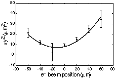
Another measure of the system performance is a scan
of the laser waist, shown in figure 5. In this scan, the e+/-
beam is used to measure the divergence of the laser by making
a series of ss
measurements as it is steered along the laser path. In the waist
scan, the effective divergence of the laser IP can be determined.
The phase space volume of the laser beam cannot be determined
from this scan since the contribution of the electrons to the
minimum measured width is not accurately known.
We would like to acknowledge support for this project
from the SLD collaboration, D. Burke and J. Sheppard. N. Phinney,
P. Raimondi and F. Zimmermann helped with data acquisition and
interpretation. Technical support from K. Ratcliffe, C. Rago,
O. Millican, A. Farvid H. Baruz, K. Underwood and L. Hendrickson
is also greatly appreciated. D. Meyerhofer of the University of
Rochester gave much appreciated review and technical assistance
during the course of the project.